
Module 14
The heart and circulation
How a simple beating tube becomes a four-chambered heart, when the heartbeat starts and what a scan shows, and how the circulation changes with a baby's first breath.
- 4lessons
- 14 minreading
- 6quiz questions
The lessons and the quiz in this module are part of the Fertility with Kara Package. Module 1 is free to read.
Your way through Module 14
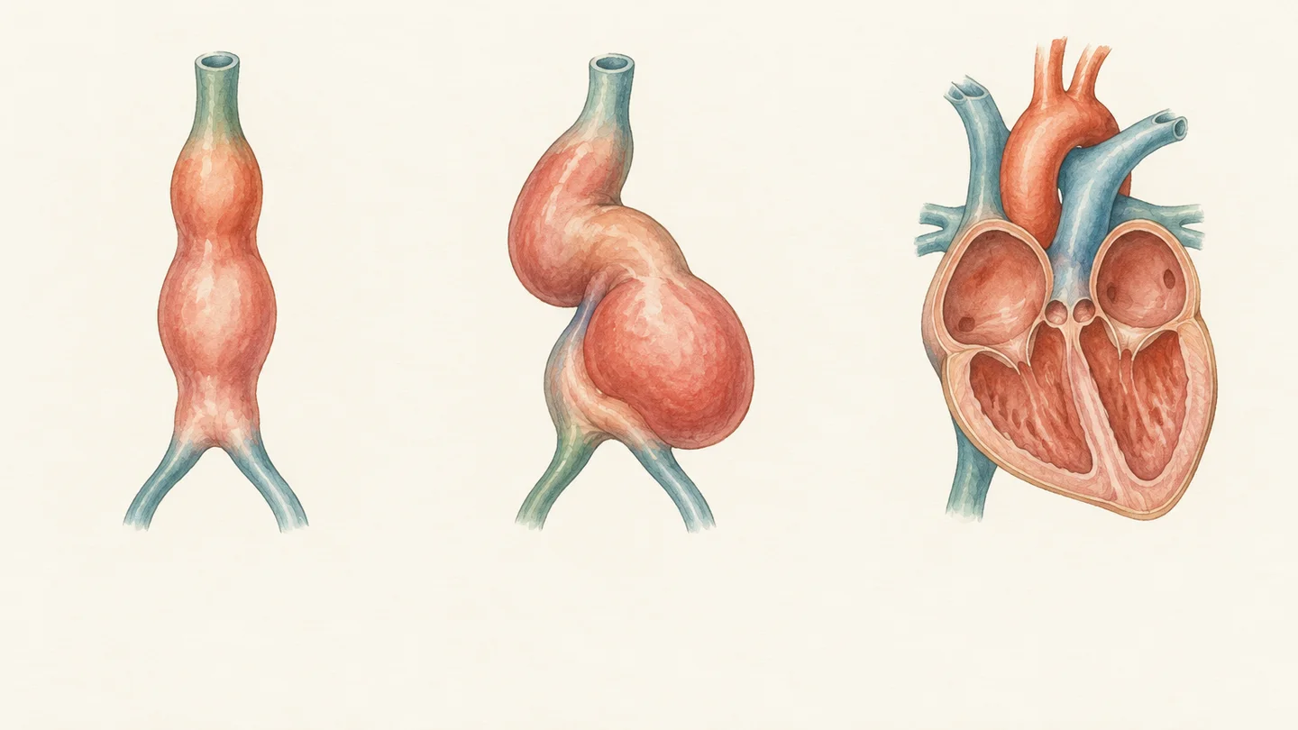
- Lesson 14.1 · 3 min Start hereRead From a tube to four chambers How the heart starts as a simple beating tube, loops into shape and is divided into four chambers, all without stopping.
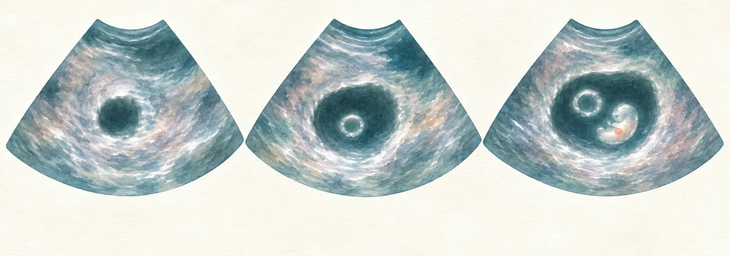
- Lesson 14.2 · 4 min Start hereRead When does the heart start beating? When the first heartbeats happen, when an early scan can see them, and how the heart rate changes in the first weeks.
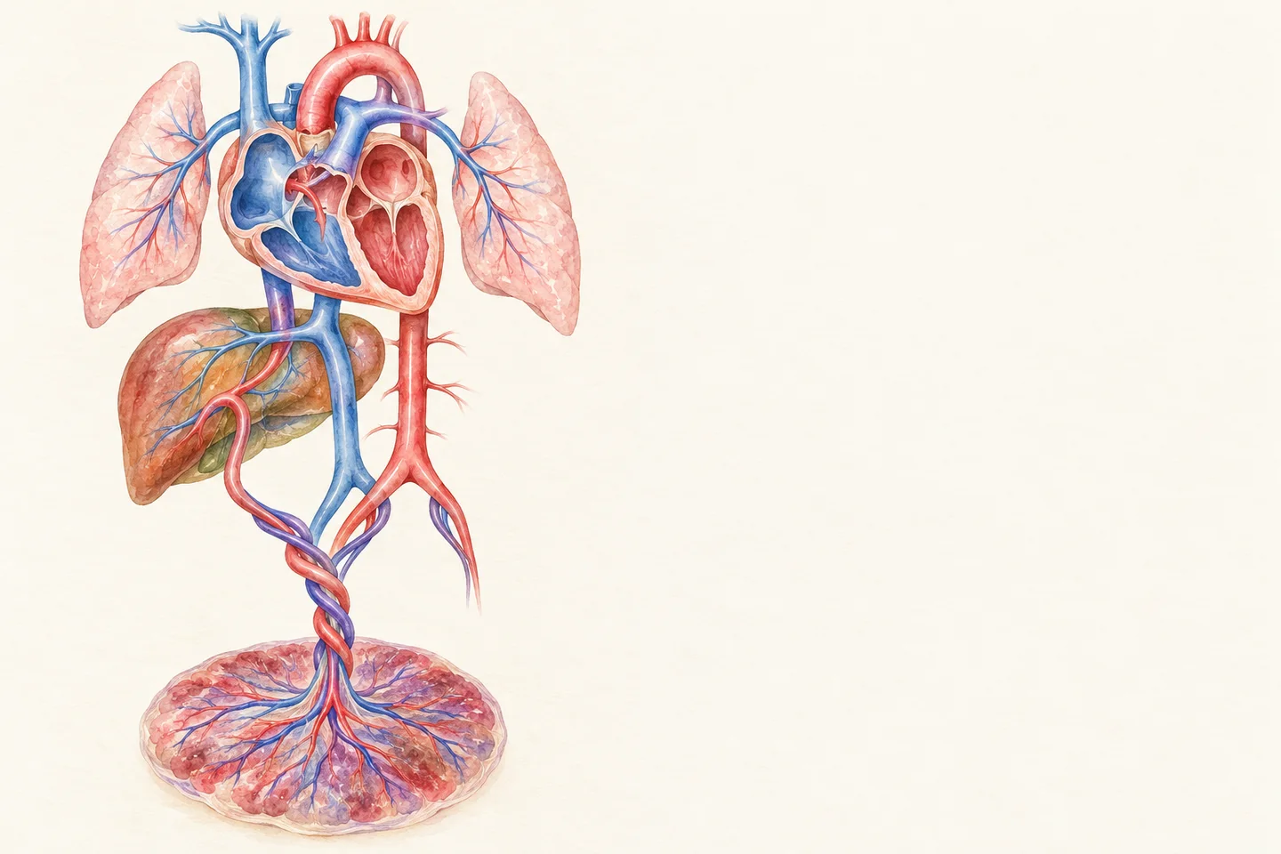
- Lesson 14.3 · 3 min Start hereRead Circulation before and after birth Why a baby's blood takes three shortcuts before birth, and how they close within minutes, days and weeks of the first breath.
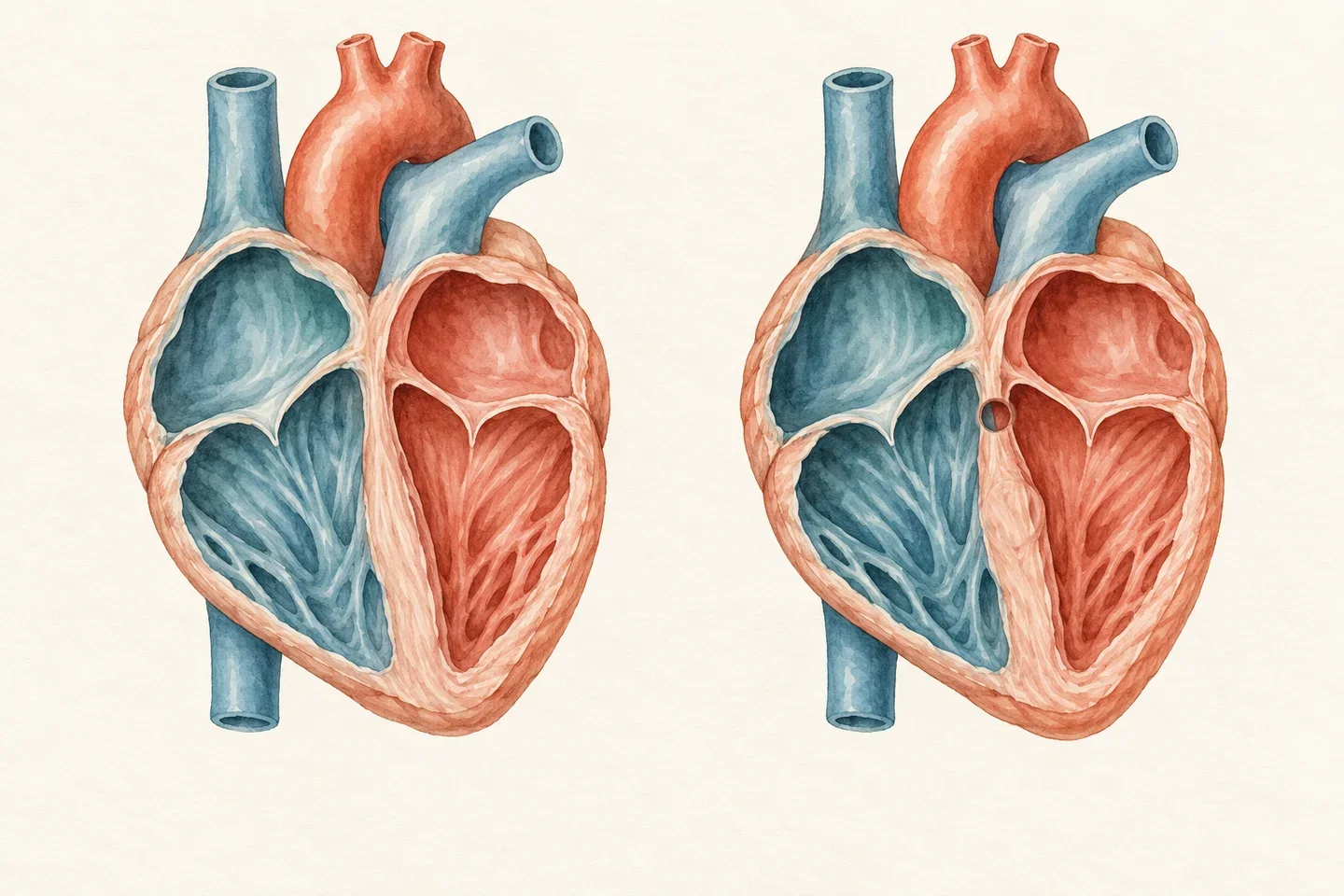
- Lesson 14.4 · 4 min Start hereRead When the heart forms differently The most common heart differences in general terms, why the heart is vulnerable while it forms, and how differences are found.

Learning aims
By the end of this module you will be able to:
- Describe how the heart tube forms, loops and is divided into four chambers.
- Explain when the heart starts beating and what an early scan can show.
- Describe the three shortcuts in the circulation before birth and what each one bypasses.
- Explain how the first breaths close the shortcuts, and over what timescale.
- Recognise the common heart differences in general terms, and compare screening and diagnosis.
- Ask your clinic sensible questions about the timing of the first scan and about heart checks.
The module in brief
The module in brief
- The heart forms as a tube from mesoderm in week 3 after fertilisation and starts to beat at about day 22, about 5 weeks of pregnancy.
- In week 4 the tube loops to the right, and walls divide it into four chambers by the end of week 7 to 8; a spiralling wall splits the outflow into the aorta and the pulmonary trunk.
- On a transvaginal scan the heartbeat is usually first seen at about 5 and a half to 6 and a half weeks of pregnancy, at about 100 beats a minute, rising to about 175 at around 8 and a half weeks.
- Before birth, the ductus venosus, foramen ovale and ductus arteriosus route oxygen-rich blood from the placenta past the liver and the fluid-filled lungs.
- At birth the lungs open, the foramen ovale is pressed shut within minutes to hours, and the ductus arteriosus closes within 1 to 4 days and is sealed for good over the following weeks, usually by 3 months.
- About 9 in 1,000 babies are born with a heart difference; most have no known cause, and many are minor.
- The detailed mid-pregnancy scan is a screening look at the heart; a specialist heart scan can give a diagnosis.