
Module 18
The kidneys and bladder
How the embryo builds three kidneys to keep one, why the baby's urine matters so much for the amniotic fluid and the lungs, and how the kidneys are linked to the reproductive organs.
- 4lessons
- 15 minreading
- 6quiz questions
The lessons and the quiz in this module are part of the Fertility with Kara Package. Module 1 is free to read.
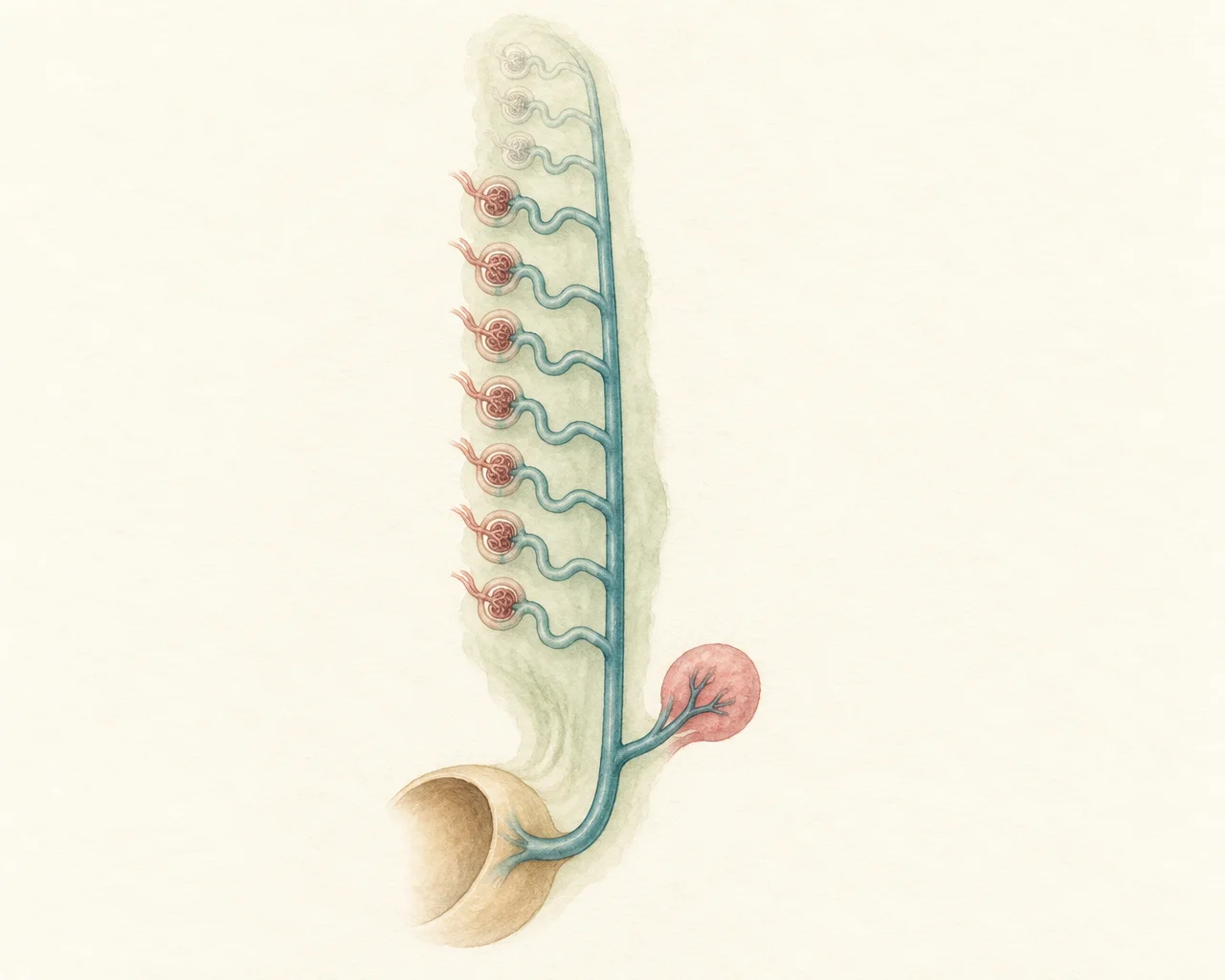
Your way through Module 18
- Lesson 18.1 · 4 min Start hereRead Three kidneys, one after another Why the embryo builds three sets of kidneys in a row, what each one does, and which one you keep.
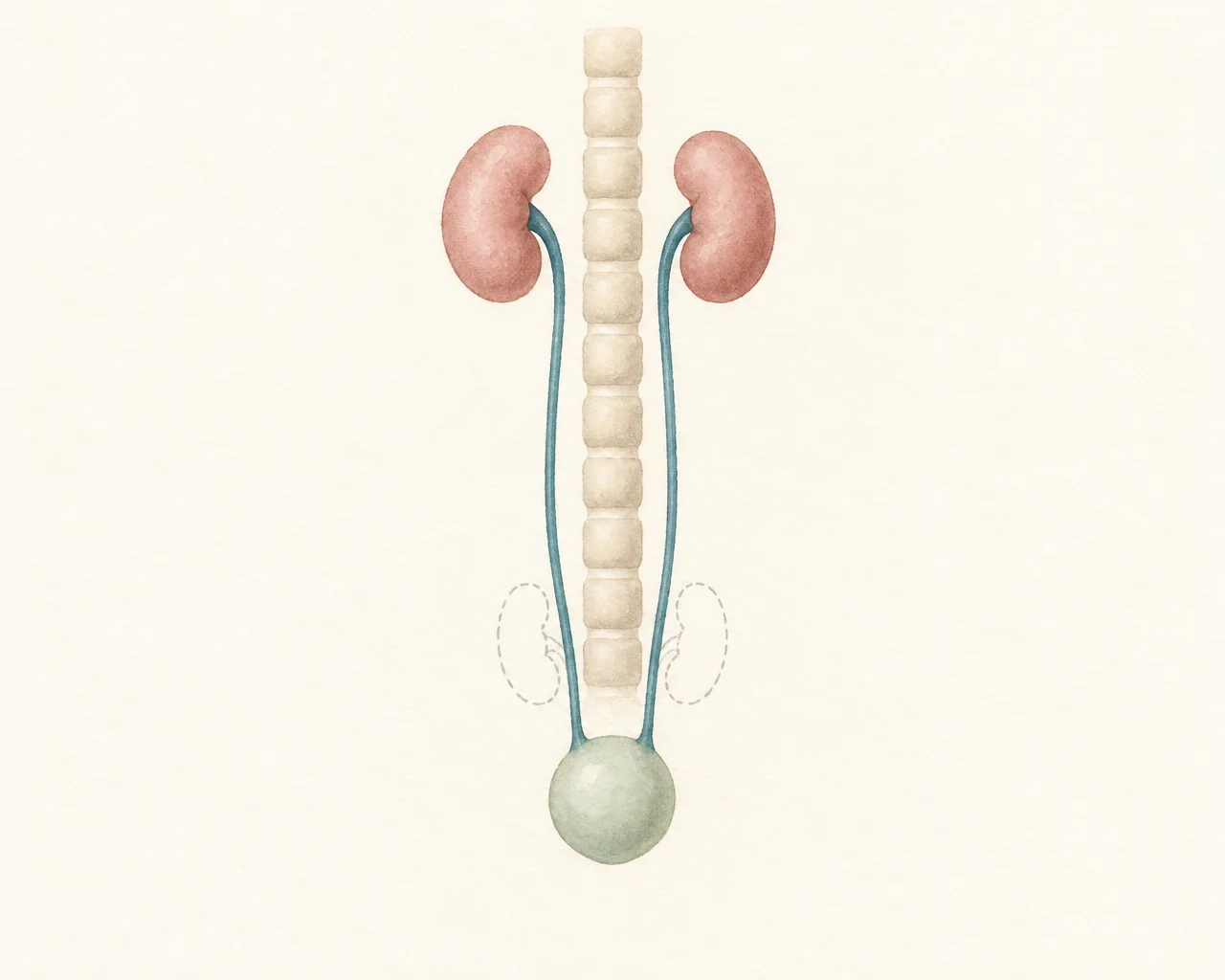
- Lesson 18.2 · 4 min Start hereRead How the kidney builds itself and rises How the ureteric bud and its cap of tissue build the kidney's plumbing and filters together, and why the kidneys seem to climb out of the pelvis.
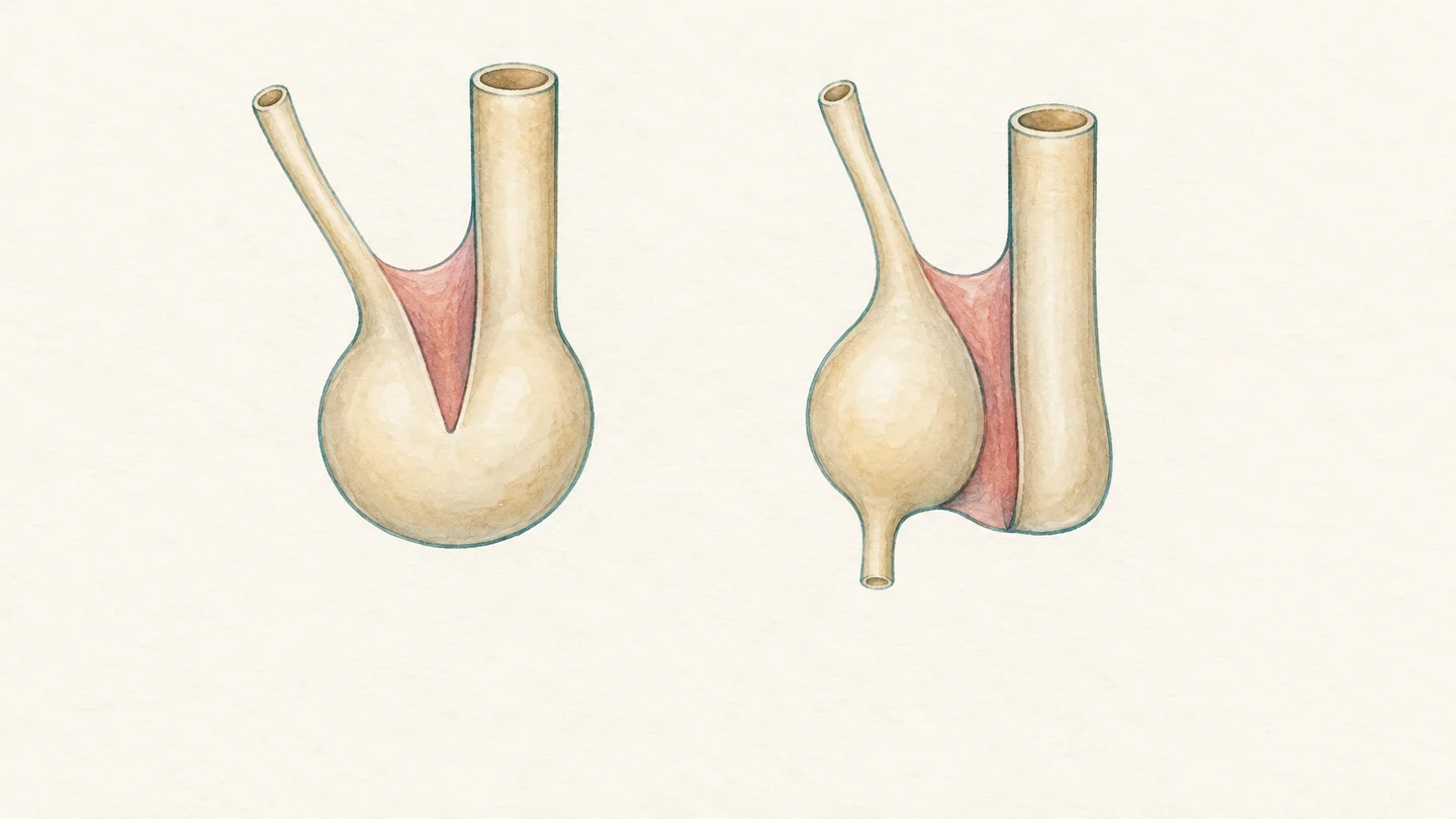
- Lesson 18.3 · 3 min Start hereRead The bladder, the baby's urine and the amniotic fluid How the bladder forms from the end of the gut, and why a baby's urine matters so much for the fluid around it and for its lungs.
- Lesson 18.4 · 4 min Start hereRead Kidneys and the reproductive system Why kidney differences and reproductive differences often turn up together, and what the common kidney findings on a pregnancy scan mean.

Learning aims
By the end of this module you will be able to:
- Describe the three generations of kidney and explain which one becomes the permanent kidney.
- Explain how the ureteric bud and the surrounding tissue build the kidney together, and why the kidneys rise.
- Describe how the cloaca, one shared opening, is split in two to form the bladder and the rectum.
- Explain why fetal urine is the main source of amniotic fluid and why that matters for the lungs.
- Recognise why kidney differences and reproductive differences can occur together.
- Ask informed questions about kidney findings on a pregnancy scan.
The module in brief
The module in brief
- The embryo builds three kidneys in a row: the pronephros, which never works, the mesonephros, which works for a few weeks, and the metanephros, the permanent kidney from week 5 after fertilisation.
- The ureteric bud and the metanephric mesenchyme build the kidney together by signalling to each other: the bud forms the pipes and the mesenchyme forms the filters.
- All nephrons are made before birth, by about 36 weeks of pregnancy, and the normal number per kidney varies widely.
- The kidneys start in the pelvis and come to lie beside the spine by week 9 after fertilisation, mainly because the body below them grows.
- One shared opening, the cloaca, is split in two by the urorectal septum: the bladder forms at the front and the rectum at the back.
- Before birth the placenta removes waste; fetal urine is the main source of amniotic fluid from about 20 weeks of pregnancy, and the lungs need that fluid to grow.
- The kidneys and the reproductive organs share an origin, so a missing kidney can go with a uterine difference in women or a missing vas deferens in men.
- The most common kidney finding on a pregnancy scan is mild urinary tract dilation, which is usually followed up with another scan.